Digital Motion X-ray (DMX) is a revolutionary diagnostic tool that is used to analyze and diagnose movement-related disorders. This technology has revolutionized the medical field and has enabled doctors and radiologists to diagnose conditions that were previously difficult to diagnose. DMX is a type of medical imaging technology that enables radiologists to capture a series of X-ray images of the patient while they are in motion. This technology provides accurate and precise diagnosis of conditions that involve movement, such as musculoskeletal disorders, sports injuries, and motor vehicle accidents. This blog post will provide an overview of Digital Motion X-ray technology, its applications, how it is performed, interpretation of the results, advantages, and limitations.
What is Digital Motion X-ray?
Digital Motion X-ray is a type of medical imaging technology that enables radiologists to capture a series of X-ray images of the patient while they are in motion. This technology is different from traditional X-rays, which only provide static images of the patient. Digital Motion X-ray technology uses digital sensors and a computer to create a video of the patient’s movement. The video can then be slowed down, analyzed, and used to diagnose movement-related conditions.
Compared to other imaging modalities, DMX technology is superior in its ability to capture the precise location and movement of bones and joints in real-time. This makes it an essential diagnostic tool for orthopedic and sports medicine physicians, chiropractors, physical therapists, and other medical professionals.
Benefits of using DMX for diagnosis and treatment planning:
The benefits of using DMX for diagnosis and treatment planning are many. Some of the benefits include:
- Accurate diagnosis: DMX technology provides a more accurate diagnosis of conditions that involve movement. This technology enables doctors to capture the precise location and movement of bones and joints in real-time, enabling a more accurate diagnosis.
- Improved treatment planning: DMX technology enables doctors to create a treatment plan that is tailored to the patient’s specific condition. This technology allows doctors to see the precise location and movement of bones and joints, enabling them to create a more effective treatment plan.
- Better patient outcomes: DMX technology enables doctors to diagnose and treat conditions more accurately, leading to better patient outcomes.
Applications of Digital Motion X-ray
Digital Motion X-ray has a wide range of applications in the medical field. Some of the applications of DMX technology include:
- Musculoskeletal disorders: DMX technology is used to diagnose and treat a wide range of musculoskeletal disorders. This includes conditions such as herniated discs, arthritis, and scoliosis.
- Sports injuries and athletic performance analysis: DMX technology is used to diagnose and treat sports injuries, such as sprains, strains, and fractures. It is also used to analyze and improve athletic performance.
- Motor vehicle accident (MVA) evaluations: DMX technology is used to diagnose and treat injuries sustained in motor vehicle accidents. This includes injuries such as whiplash and spinal cord injuries.
- Industrial and occupational injuries: DMX technology is used to diagnose and treat injuries sustained in the workplace. This includes injuries such as repetitive stress injuries and spinal cord injuries.
How Digital Motion X-ray is performed
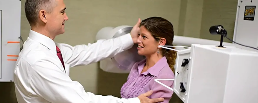
The DMX examination is performed in a radiology suite by a trained radiologic technologist under the supervision of a radiologist. The patient is positioned on an exam table, and the X-ray machine is positioned above them. The patient is then instructed to perform the movement that needs to be examined, such as bending over, twisting, or lifting. The X-ray machine captures a series of images of the patient while they are in motion.
Patient preparation and safety precautions for Digital Motion X-Ray:
Before the DMX examination, the patient is required to remove any jewelry or
other metal objects that could interfere with the X-ray machine. The patient is also required to wear a lead apron to protect them from unnecessary radiation exposure. The radiologic technologist will then provide instructions to the patient on the movement that needs to be examined.
Equipment and technology used in DMX:
DMX technology requires specialized equipment and technology to capture the images of the patient while they are in motion. The equipment used in DMX includes:
- X-ray machine: DMX technology requires an X-ray machine that is capable of capturing a series of images of the patient while they are in motion. This X-ray machine is usually mounted on a track or a stand that enables it to move with the patient.
- Digital sensors: DMX technology uses digital sensors that are placed on the patient’s skin to capture the movement of bones and joints. These sensors are connected to a computer that processes the data and creates a video of the patient’s movement.
Steps involved in DMX examination:
The DMX examination involves several steps, which include:
- Patient positioning: The patient is positioned on an exam table in a way that allows the X-ray machine to capture the movement that needs to be examined.
- X-ray machine positioning: The X-ray machine is positioned above the patient in a way that allows it to capture a series of images of the patient while they are in motion.
- Movement instructions: The radiologic technologist provides instructions to the patient on the movement that needs to be examined.
- Image capture: The X-ray machine captures a series of images of the patient while they are in motion.
- Data processing: The data captured by the digital sensors is processed by a computer to create a video of the patient’s movement.
- Interpretation: The radiologist interprets the video and provides a diagnosis of the patient’s condition.
Interpretation of Digital Motion X-ray
Interpreting DMX results requires specialized training and expertise. The interpretation of DMX results is usually done by a radiologist or a physician who has received specialized training in interpreting DMX results.
Role of radiologists and physicians in DMX interpretation:
Radiologists and physicians play a critical role in interpreting DMX results. These professionals use specialized software and techniques to analyze the video and determine the patient’s condition. The interpretation of DMX results requires a high level of expertise and training, as the interpretation is critical for successful treatment outcomes.
Techniques and tools used for analysis and measurement:
Radiologists and physicians use specialized software and techniques to analyze and measure the patient’s movement in the DMX video. Some of the techniques and tools used for analysis and measurement include:
- Angle measurement: Radiologists and physicians use specialized software to measure the angles between bones and joints in the DMX video. This helps them determine the patient’s range of motion and identify any abnormalities.
- Distance measurement: Radiologists and physicians use specialized software to measure the distance between bones and joints in the DMX video.
- Frame-by-Frame Analysis: The video captured by the Digital Motion X-Ray is analyzed frame by frame to identify any abnormalities or conditions. This allows for a more detailed examination of the motion and movement of the joints and soft tissues.
- Advanced Software: Advanced software is used to process the video and create a high-quality image that can be analyzed more easily. This software allows the medical professional to manipulate and zoom in on specific areas of the video for a closer examination.
- Comparison to Normal Range of Motion: The interpretation of Digital Motion X-Ray also involves comparing the patient’s range of motion to the normal range of motion for the affected joint or area. This allows the medical professional to identify any limitations or abnormalities that may be present.
- Clinical Correlation: Digital Motion X-Ray interpretation is often done in conjunction with other diagnostic tools and tests to provide a more complete picture of the patient’s condition. Medical professionals use their clinical judgment to correlate the findings from the Digital Motion X-Ray with other diagnostic tests and the patient’s symptoms to provide an accurate diagnosis.
How much radiation exposure is there with DMX?
The table below shows annual occupational dose limits recommended by the Code of Federal Regulations in the United States for adults to be exposed to are 5000 mrem for the whole body.
Common Radiation Doses in mrem.
| Source | Amount of Exposure (mrem)1-10 |
|---|
| MRI or ultrasound | 0 |
| X-ray inspect at airport | .002 |
| Airplane travel/per hour | 0.5 |
| One static Cervical image with DMX | 7 |
| ½ pack of cigarettes smoked | 18 |
| Standard chest x-ray | 20 |
| One standard static hip x-ray | 70-120 |
| One standard static X-ray of cervical spine | 70-120 |
| DMX full exam of peripheral joint | 150-350 |
| Complete lumbar/cervical standard x-ray series | 600-800 |
| Average annual exposure in US | 620 |
| Complete DMX neck exam (90 seconds) | 550* |
| Normal Fluoroscopy (non-DMX machine) | 750 |
| (90 seconds of continuous exposure) | |
| CT of spine | 1000 |
| High dose Fluoroscopy (non-DMX machine) | 1500 |
| (90 seconds of exposure) | |
*Approximately 550 mrem….the actual DMX machine measures total radiation exposure with each scan. Some scans at 90 seconds are more and some less than this depending on the collimation of the beam (filtering and amount of site exposure). Scans that are less than 90 seconds will typically have less than 550 mrem and those that are more than 90 seconds more than this number.
Is DMX Good For My Condition(s)?
This type of imaging can help diagnose a wide range of conditions affecting the bones, joints, and soft tissues, including:
- Joint instability: Digital Motion X-Ray can help identify any instability or abnormal movement of the joints. This can be particularly useful in diagnosing conditions such as shoulder instability, knee instability, and spinal instability.
- Ligament injuries: Ligament injuries are common in sports and can be difficult to diagnose with traditional imaging techniques. Digital Motion X-Ray allows medical professionals to see the movement of the ligaments in real-time, which can help identify any tears or injuries.
- Spinal misalignment: Digital Motion X-Ray can help diagnose conditions such as scoliosis, which involve a curvature or misalignment of the spine.
- Conditions where regenerative therapies may be an alternative to surgery. A high percentage of the case we see at Joint and Arthritis Pain Relief have been told surgery is their only option. After receiving a DMX it may point them to more conservative options such as PRP injections.
- Osteoarthritis: Digital Motion X-Ray can help identify any joint damage or degeneration caused by osteoarthritis.
- Post-surgical evaluation: Digital Motion X-Ray can help evaluate the effectiveness of surgical treatments and rehabilitation programs by assessing the movement and stability of the affected joint or area.
Digital Motion X-Ray can be a valuable tool in the diagnosis of musculoskeletal conditions. Particularly those that involve movement and motion. Patients can use this to their advantage when trying to find the best treatment option for their knee, back, hip, or neck pain. It provides a more comprehensive picture of what’s happening inside the body and can help medical professionals develop a more effective treatment plan for patients.
Digital Motion X-Ray Summary
Digital Motion X-Ray is a revolutionary diagnostic imaging technology that provides a more comprehensive picture of what’s happening inside the body. By capturing motion and movement in real-time, it allows for a more accurate diagnosis of various musculoskeletal conditions. Compared to traditional x-ray imaging, Digital Motion X-Ray provides several advantages. This includes the ability to diagnose conditions that may be missed with traditional imaging. It also allows the ability to assess the effectiveness of various treatments and rehabilitation programs.
With its applications in sports medicine, auto accidents, and other severe injuries, Digital Motion X-Ray is a valuable tool for physicians and patients alike. It offers a cost-effective, non-invasive, and efficient way to diagnose various conditions and evaluate the progress of treatment. Search Digital Motion X-Ray near me to find a clinic that offers the service.
As the technology behind Digital Motion X-Ray continues to improve, it’s likely that its applications will expand even further, allowing for more accurate and comprehensive diagnoses of various conditions. Overall, Digital Motion X-Ray represents a significant step forward in the field of diagnostic imaging and has the potential to transform the way we diagnose and treat musculoskeletal conditions.
If you have been told that surgery is your only option, seek a second opinion today.
Learn More About Alternative Ways to Alleviate Your Pain
Get back to doing the things you love, faster & without surgery.
Request an Appointment Today!